
Module 1: Vascular Ultrasound
Lesson 1: Microcirculation, Blood Flow Pathway, and Vessel Walls
Overview
Microcirculation plays a critical role in oxygen exchange, nutrient delivery, and waste removal at the capillary level. Understanding the structure of blood vessels and the flow pathway is essential for vascular sonographers to evaluate normal vascular anatomy and function.
This lesson will cover:
The microcirculation system (arterioles, capillaries, venules)
The blood flow pathway from arteries to veins
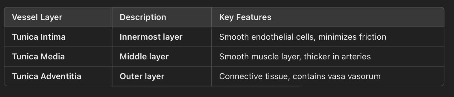
The three layers of vessel walls and their functions
1. Blood Flow Pathway and Microcirculation
Blood moves through a network of progressively smaller and larger vessels, following this general sequence:
Artery → Arteriole → Capillary → Venule → Vein
🔹 Arteries
Carry oxygenated blood away from the heart (except pulmonary arteries).
Have thicker muscular walls to withstand high pressure.
Progressively branch into smaller arteries before becoming arterioles.
🔹 Arterioles (Resistance Vessels)
Control blood pressure and flow distribution via vasoconstriction and vasodilation.
Act as the primary site of vascular resistance, regulating perfusion to tissues.
🔹 Capillaries
Smallest blood vessels, one cell thick to allow for gas and nutrient exchange.
Transition point between the arterial and venous systems.
🔹 Venules
Small veins that collect deoxygenated blood from capillaries and transport it to larger veins.
Have thinner walls than arterioles.
🔹 Veins
Carry deoxygenated blood back to the heart (except pulmonary veins).
Contain valves to prevent backflow, especially in the lower extremities.
Progressively increase in size as they return blood toward the heart.
2. Vessel Wall Layers: Arteries vs. Veins
Each vessel is composed of three layers that contribute to its function.
Note: Tunica adventitia means the same as tunica externa.
Note: Tunica adventitia/externa contains the vasa vasorum.
Vasa vasorum has a latin meaning of vessels of the vessels.
Vasa Vasorum are tiny vessels that supply the walls of larger arteries and veins.
Arteries vs. Veins: Structural Differences
Arteries:
Thicker tunica media → helps maintain blood pressure.
More elastic fibers → accommodate pulsatile flow.
No valves → flow is pressure-driven.
Veins:
Thinner tunica media → lower resistance, collapsible walls.
Contain valves → prevent backflow, especially in the legs.
More distensible → can expand to hold more blood.
3. Clinical Examples
Example 1: Arterioles and Blood Pressure Regulation
A patient presents with hypertension, and Doppler ultrasound shows increased resistance in peripheral arteries.
This suggests that the arterioles are constricted, increasing vascular resistance and elevating blood pressure.
Example 2: Venous Valves and Chronic Venous Insufficiency (CVI)
A patient complains of leg swelling and varicose veins.
Duplex ultrasound reveals valve incompetence in the great saphenous vein, leading to venous reflux.
This supports a diagnosis of chronic venous insufficiency, where blood pools due to ineffective valve function.
Summary
Blood flows from arteries → arterioles → capillaries → venules → veins.
Arterioles regulate blood pressure and control microcirculation.
Capillaries facilitate gas exchange and nutrient transfer.
Veins have thinner walls and valves to assist venous return.
The three vessel layers (intima, media, adventitia/externa) contribute to structure and function.